


Projekte der Abteilung Funktionelle Neuroanatomie
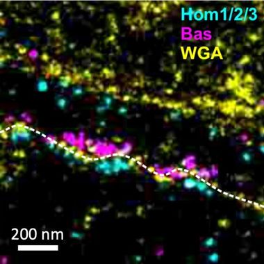
Molecular Nanoarchitecture of the Synapse
Molecular Nanoarchitecture of the Synapse
The synapse, both its pre- and postsynaptic compartments, can be considered a complex assembly of interacting proteins that need to be tightly regulated in space and time. Much like real machines, all the components of these molecular machines need to be at their place to enable the highly specific and extraordinarily rapid process of synaptic transmission. In collaboration with Prof. Dr. Mike Heilemann, we developed maS3TORM, a fully automated highly multiplexed super-resolution microscope that allows us to identify up to 20 proteins in the same synapse simultaneously in 3D at nanometer resolution. Using this approach at the calyx of Held and cultured neurons, we aim at deciphering the nanostructural principles that underly the molecular design of synapses.
Venkataramani et al. (2016), Nature Methods; Klevanski et al. (2020), Nature Communications.
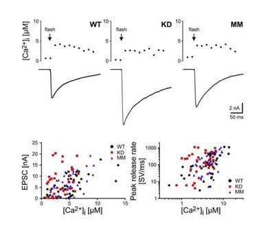
Structure and Function of Synapses
To reveal the contribution of presynaptic proteins in defining fundamental properties of synaptic transmission, we use the calyx of Held excitatory model synapse that allows direct pre- and postsynaptic electrophysiological and optical recordings. Molecular perturbations are targeted to the calyx by stereotactic delivery to the cochlear nucleus. In conjunction with these techniques, 3D volume scanning electron microscopy and super-resolution microscopy allow detailed studies of structure-function relationships.
Körber et al. (2015), Neuron; Parthier et al. (2018), J Physiol.
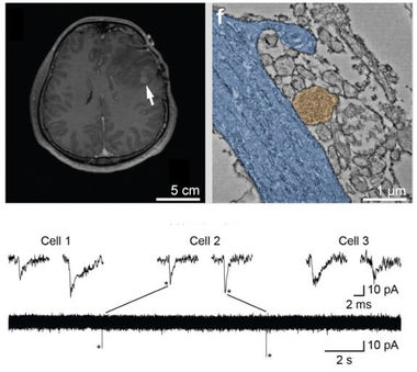
Synapses between Neurons and Tumors
In 2019 we discovered a new category of synapses, formed between neurons and glioblastoma cells: the neurogliomic synapse. As we could demonstrate in collaboration with Prof. Dr. Frank Winkler, neurogliomal synapses directly drive tumor progression by relaying neuronal activity to tumor cells. Neurogliomal synapses utilize Ca2+ permeable glutamate receptors of the AMPA subtype and trigger postsynaptic EPSCs and Ca2+ signals, which in turn affect proliferation and migration of tumor cells. Using a highly multidisciplinary approach, we now aim at understanding the signalling mechanisms involved in this special type of synaptic communication. Furthermore, we aim at establishing “neurotumoral” synapses as general principle of communication between neurons and tumors. Venkataramani et al. (2019), Nature;
Venkataramani et al. (2020), Neuro-Oncology.
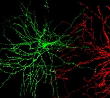
Neuronal Structural Plasticity in Chronic Pain
Chronic pain is a percept generated in the neocortex. One possible mechanism of chronification could be structural plasticity of cortical neurons. Chronic pain induces changes in grey matter volume, both in humans and rodents, and points towards structural changes on the cellular level. We study this by comparing MRI and two-photon in vivo imaging in a longitudinal study design. Also, we repetitively image entire cortical neurons and their structural changes upon induction of chronic pain, allowing us to correlate synaptic and dendritic structural plasticity with pain chronification.
Kuner & Kuner (2020), Physiological Reviews; Asan et al. (2021), Scientific Reports.
Thalamocortical Interactions and their Behavioral Function
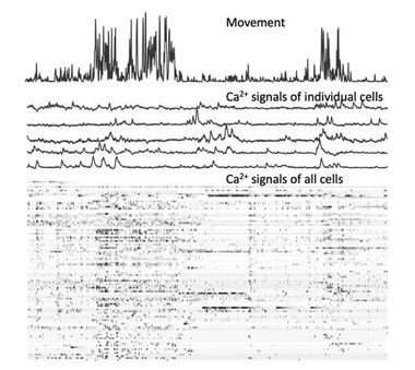
The neorcortex is vastly connected within itself, but cortical regions also communicate via the thalamus. Massive corticothalamocortical loops mediate layer 5b outputs of cortex to the thalamus, where a special type of giant synapse forwards this information onto thalamic relay cells, which in turn target cortical layers. The physiological relevance of this communication stream is so far unknown. We use head-mounted miniature cameras in combination with GRIN lenses to image the network activity of thalamic relay neurons while mice perform behavioral tasks. In collaboration with Prof. Dr. Alexander Groh, this will allow us to identify neuronal ensemble activity in relation to behavior, and hence may ultimately reveal the physiological function of corticothalamocortical loops.
Seol and Kuner (2015), European Journal of Neuroscience; Pelzer et al. (2017), Frontiers in Synaptic Neuroscience.
Synaptic Basis of Odor Discrimination Behavior
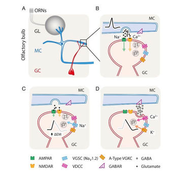
The olfactory bulb forms the first electrical representation of the odor environment and uses intricate inhibitory networks for “preprocessing” of the odor-induced activity patterns. We combine highly targeted molecular perturbations of granule cells with functional and behavioral characterizations. In collaboration with Prof. Dr. Andreas Schaefer we built a fully automated olfactometer that allows group-housed mice to perform olfactory discrimination tasks. This approach allows us to understand the neuronal mechanisms of odor discrimination behavior on a molecular and cellular level.
Nunes and Kuner (2015), Nature Communications; Nunes and Kuner (2018), PLoS Biology; Reinert et al. (2019), Frontiers in Behavioral Neurosciences; Egger and Kuner (2021), Cell and Tissue Research.