
Neuroanatomie - Skutella Gruppe

Germ stem cell reprogramming
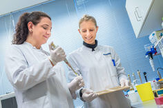
It has been suggested that the generation of human germline stem cells from primordial germ cells (PGCs) or spermatogonial stem cells (SSCs) may provide simple and non-controversial access to individual cell-based therapy. These human studies were based on mouse models of germ stem cell reprogramming, in which the molecular mechanisms underlying the shift to pluripotency could be investigated in more detail. However, recent studies revealed the impossibility of a conversion of adult mouse SSCs and human PGCs into a truly pluripotent state. For the conversion of PGCs it appeared to be necessary to conduct a two-factor induction of pluripotency with Oct4 and Sox2. In our present research, adult human germ stem cell cultures are grown from the CD49f MACS- and matrix-selected fractional part of enriched human adult spermatogonia. When compared to authentic pluripotent stem cells, these human adult germ stem cell (haGSC) cultures are not fully reprogrammed. For this reason, an investigation of the molecular blockages of pluripotency in germ cells is conducted. It would be deeply interesting to improve culture conditions and to apply bolting factors, such as small molecules that deter from the molecular block and hinder a full germ stem cell (GSC) or haGSC conversion to fully molecular pluripotent stem cells. It would also be of great interest to enlarge the restricted time window of SSC to pluripotent stem cell (PSC) conversion.
Oocyte maturation
In a cooperation project with Prof. Irma Virant-Klun from the University Hospital in Ljubljana, Slovenia, we are researching human oocytes. In IVF patients, there is a proportion of about 15-20% of immature follicles called germinal vesicles (GV). So far, these germinal vesicles could not be fertilized and are therefore discarded. However, a new method of, so-called, ‘in vitro maturation’, IVM for short, allows the follicle progenitor cells to be artificially brought to maturity in the Petri dish. We were able to show that it is possible to improve the in vitro process of human oocyte maturation in a co-culture with cumulus cells of mature oocytes of the same female in order to generate a clinically more successful and safe future procedure. This procedure can also be used for cancer patients undergoing chemotherapy or radiotherapy to maintain their fertility. In these cases, the material is removed and artificially matured before the therapy. It is no longer necessary, as before, for patients to wait valuable time until, by in vivo stimulation, mature oocytes can be obtained. The research groups of Prof Skutella and Virant Klun are currently working to better understand the rate of oocyte maturation in gonadotropins (FSH, hCG and AMH) in relation to the age of oocyte donors at the single cell level. They study the expression of transcription factors previously identified in a pilot study of human oocytes in vitro maturation, including the oocyte-specific genes FIGLA and H1FOO in individual in vitro matured oocytes. Furthermore, the research groups analyze the effect of gonadotropins and the female age on the regulation mechanisms of miRNAs on the expression of transcription factors and other important identified genes in individual oocytes, comparing the exosomal miRNA of in vivo and in vitro matured oocytes. They expect that the results will provide an improved future understanding of the molecular basis of female fertility and may provide a basis for the potential development of new drugs and therapies for ovarian hormone stimulation in IVM.
References
Virant-Klun I, Bauer C, Ståhlberg A, Kubista M, Skutella T. Human oocyte maturation in vitro is improved by co-culture with cumulus cells from mature oocytes. Reprod Biomed Online. 2018 May;36(5):508-523. doi: 10.1016/j.rbmo.2018.01.011. Epub 2018 Feb 5. PubMed PMID: 29503212.
Conrad S, Azizi H, Skutella T. Single-Cell Expression Profiling and Proteomics of Primordial Germ Cells, Spermatogonial Stem Cells, Adult Germ Stem Cells, and Oocytes. Adv Exp Med Biol. 2018;1083:77-87. doi: 10.1007/5584_2017_117. PubMed PMID: 29299873.
Virant-Klun I, Ståhlberg A, Kubista M, Skutella T. MicroRNAs: From Female Fertility, Germ Cells, and Stem Cells to Cancer in Humans. Stem Cells Int. 2016;2016:3984937. doi: 10.1155/2016/3984937. Epub 2015 Nov 9. Review. PubMed PMID: 26664407; PubMed Central PMCID: PMC4655303.
Molecular atlas of human spermatogenesis
Furthermore we are working on a molecular atlas of different stages of human spermatogenesis Detailed knowledge of the molecular histological profile of the process of human spermatogenesis and the principal participating cells is an important prerequisite to develop strategies for diagnostics and therapy of male infertility and how to handle germ cells in the management of assisted reproduction. It is also essential to better understand the manifestation of testicular cancer for the development of improved and more individualized diagnostic- and therapy options in the future. At a present stage the scientific knowledge about testicular genes and proteins in the human is still insufficiently characterized in terms of cell type specific localization and function. The establishment of a more testis specific molecular atlas would be significant for the organization of our knowledge and the increase of molecular understanding of human reproductive biology and disease