
- Institut
-
Herz- und Kreislaufphysiologie
- Markus Hecker
- Thomas Korff
-
Hugo H. Marti
-
Forschung
- Zelluläre und molekulare Mechanismen der postnatalen Entwicklung des zerebralen Gefäßsystems
- Die Bedeutung der molekularen PHD-HIF Achse für den akuten Schutz und die langfristige Regeneration nach einem ischämischen Schlaganfall
- Charakterisierung und gezielte Aktivierung von NRF2-abhängigen antioxidativen Mechanismen beim akuten Schlaganfall
- Extrazelluläre Nukleinsäuren als Trigger neuroinflammatorischer Prozesse in akuten und chronisch degenerativen Erkrankungen des Zentralnervensystems
- Neuroprotektion und Neurogenese
- Blut-Hirn-Schranke
- Publikationen
- Personal
-
Forschung
- Andreas H. Wagner
- Neuro- und Sinnesphysiologie
- Lehre
- Zentrale Einrichtungen
- Bernard Katz Lecture
- Stellenangebote
- Aktuelles
Forschung
Vascular Organoids for Aminal Welfare (3R)
Differentiation of endothelial cells in vivo

Endothelial cells (EC) cover the inside of all blood vessels as a monolayer. In the adult mature vascular system EC contact pericytes (PC) or (sometimes) smooth muscle cells (SMC) through breaks in the underlying basal membrane (BM). Under homeostasis ECs located in an intact monolayer do not proliferate and are referred to as "quiescent".

Stimulatory events such as wounding of blood vessels and the following healing process activate ECs to degrade their underlying basal membrane, to migrate into the surrounding matrix, to proliferate and to establish new anastomosing networks, which were again covered by PCs or SMCs. Activated ECs are referred to as "angiogenic".
Differentiation of endothelial cells in vitro

In vitro primary ECs were usually cultured as two-dimensional flat monolayers reflecting some properties of the endothelial in vivo phenotype. Nevertheless, ECs cultured in that way are not completely quiescent. Even in a confluent monolayer up to 10% of the EC proliferate and tend to lose their differentiation over time (e.g. downregulation of CD34).

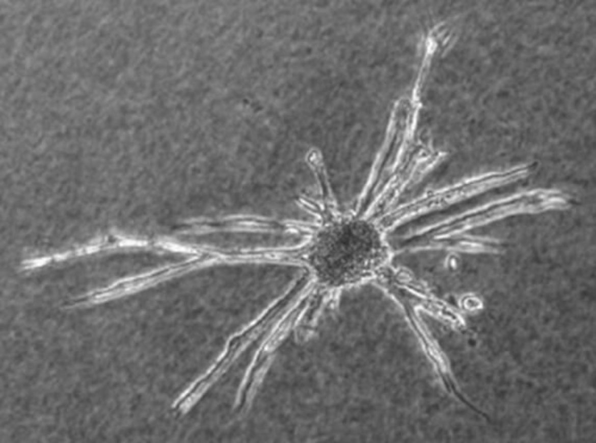
In order to preserve differentiation and quiescence of ECs in vitro, we developed a novel method to culture ECs as three-dimensional spheroids. Single suspended ECs are seeded under non-adhesive conditions in round bottom 96-well plates or hanging drops. After 18-24h, all ECs seeded in one well/drop contribute to the formation of a single spheroid. The spheroids organize over time (24 h) to establish a quiescent, non-proliferating surface monolayer of endothelial cells enclosing a core of (quiescent) unorganized cells. We exploit this cell culture technique to analyze angiogenesis and endothelial differentiation.
- Institut
-
Herz- und Kreislaufphysiologie
- Markus Hecker
- Thomas Korff
-
Hugo H. Marti
-
Forschung
- Zelluläre und molekulare Mechanismen der postnatalen Entwicklung des zerebralen Gefäßsystems
- Die Bedeutung der molekularen PHD-HIF Achse für den akuten Schutz und die langfristige Regeneration nach einem ischämischen Schlaganfall
- Charakterisierung und gezielte Aktivierung von NRF2-abhängigen antioxidativen Mechanismen beim akuten Schlaganfall
- Extrazelluläre Nukleinsäuren als Trigger neuroinflammatorischer Prozesse in akuten und chronisch degenerativen Erkrankungen des Zentralnervensystems
- Neuroprotektion und Neurogenese
- Blut-Hirn-Schranke
- Publikationen
- Personal
-
Forschung
- Andreas H. Wagner
- Neuro- und Sinnesphysiologie
- Lehre
- Zentrale Einrichtungen
- Bernard Katz Lecture
- Stellenangebote
- Aktuelles